Understanding Sciatica: What It Is, What It Isn’t, and How It’s Treated
“Sciatica” is one of the most commonly misused labels in back-pain conversations. Many people assume that any pain in the leg coming from the back must be sciatica, but that’s simply not true. Real sciatica is much less common than most people believe, accounting for only 5–10% of all low-back-pain cases. That means 90–95% of people with back pain do not have sciatica.
This article breaks down what sciatica actually is, how to recognise it, what causes it, and what current research says about treatment and exercise.
What Sciatica Really Is
Sciatica refers to pain caused by irritation or compression of the spinal nerves, typically starting in the lower back and travelling down the leg. It can involve a mix of sensory and motor symptoms.
Common sensory symptoms:
- Numbness
- Pins and needles
- A sharp, localised shooting or burning pain
Common motor symptoms:
- Reduced leg strength
- Difficulty controlling movement
- Altered posture or gait
These symptoms occur because the irritated nerve affects both how the leg feels and how it functions.
Common Misconceptions About Sciatica
Around two-thirds of people with low-back pain also report leg pain, and many assume that any leg symptom equals sciatica. However, most leg pain from the back is non-specific and does not follow a nerve pathway.
A key hallmark of true sciatica:
Pain that travels below the knee, following a predictable nerve distribution.
However, not all sciatica travels below the knee, another important indicator is that the leg pain is typically worse than the back pain when a nerve root is involved.
Why Sciatica Often Flares With Coughing or Sneezing
Coughing, sneezing, or straining increases intra-abdominal pressure, which can momentarily irritate an already affected nerve root, intensifying pain.
Where the Pain Comes From
Sciatica can originate from different lumbar spinal levels—most commonly L4–L5 or L5–S1. Each level affects a different nerve root, producing different patterns of pain, numbness, or weakness down the leg and sometimes into the foot.
The Most Common Cause: Lumbar Disc Herniation
After ruling out other conditions, about 85% of sciatica cases involve a herniated disc pressing on a nerve root.
A disc herniation is the displacement of disc material beyond the normal margins of the disc space. It occurs when the disc material, typically the softer inner material breaks through the outer ring.
The leading cause of disc herniations is genetics (Patel et al (2011) PMID: 21266637 and Battie et al (2009) PMID: 19111259). Equally there is no evidence that lifting with a bent back increases the risk of disc herniations (Swain et al (2020) PMID: 31451200 and Saraceni et al (2021) PMID: 34288926). Furthermore the spontaneous regression of herniated disc tissue can occur in between 41 and 96% of cases, and can completely resolve after conservative treatment.
Clinicians often use the straight-leg-raise test as part of assessment. A positive “crossed” straight-leg-raise test (pain in the affected leg when the opposite leg is lifted) is highly specific for nerve-root compression.
Most Sciatica Improves Naturally
The natural course is overwhelmingly positive:
- 87% of people improve within 3 months without surgery
- Motor function often recovers
- Sensory changes (e.g., numbness) may take longer but usually improve over time
First-line conservative treatment usually includes:
- Anti- inflammatory medication or other pain-relief strategies
- Guided mobility and strengthening exercise
- Avoiding prolonged bed rest and maintaining gentle activity
Epidural injections may help short-term pain but
don’t change long-term outcomes.
A healthcare professional should always guide care to ensure symptoms are managed safely.
When Should You Get an MRI or CT Scan?
Imaging is
not recommended early, because many pain-free people have disc bulges.
MRI or CT is considered when:
- There are severe or worsening neurological symptoms
- “Red flag” signs suggest infection or tumour
- Symptoms persist beyond 4–6 weeks and the patient may be a candidate for injections or surgery
What About Surgery?
Surgery may be appropriate when:
- MRI findings match the symptoms
- Pain persists despite 6+ weeks of conservative therapy
- The person prefers the option of faster relief
Most research shows:
- Surgery offers quicker relief,
- But after 1 year, outcomes are similar whether you had surgery early or stuck with conservative treatment.
Repeat surgery rates are around 6% after 1 year and 13% after 4 years.
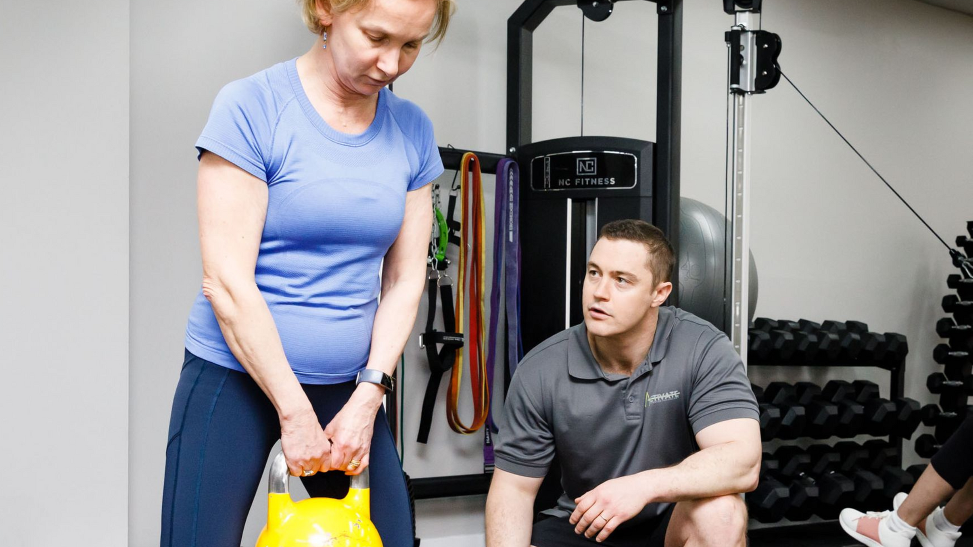
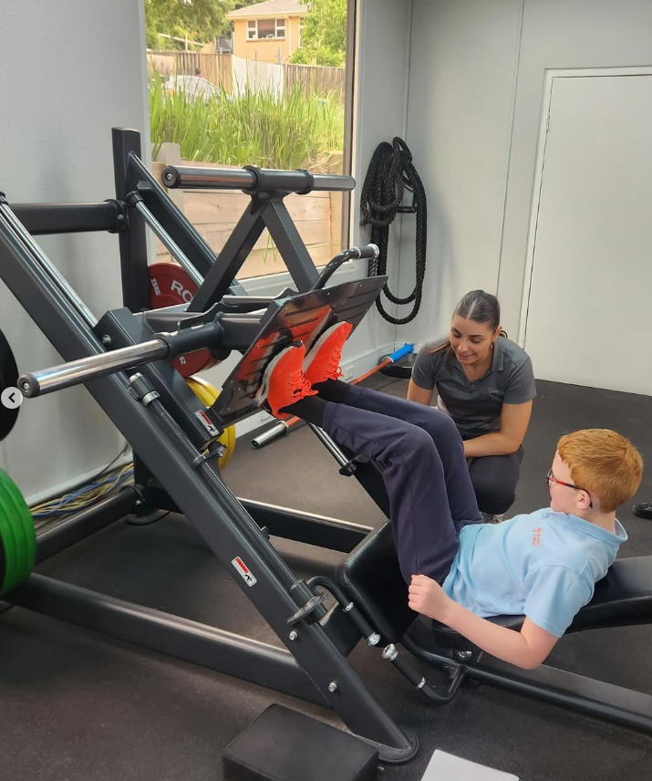
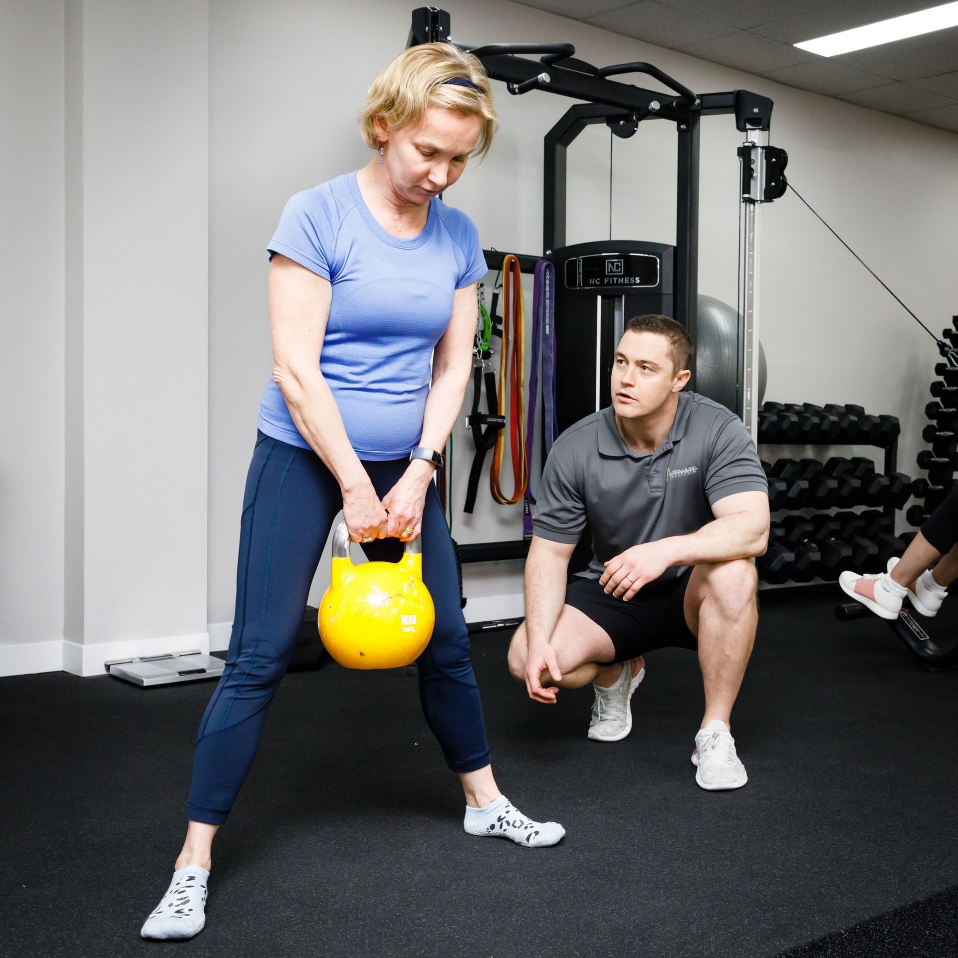
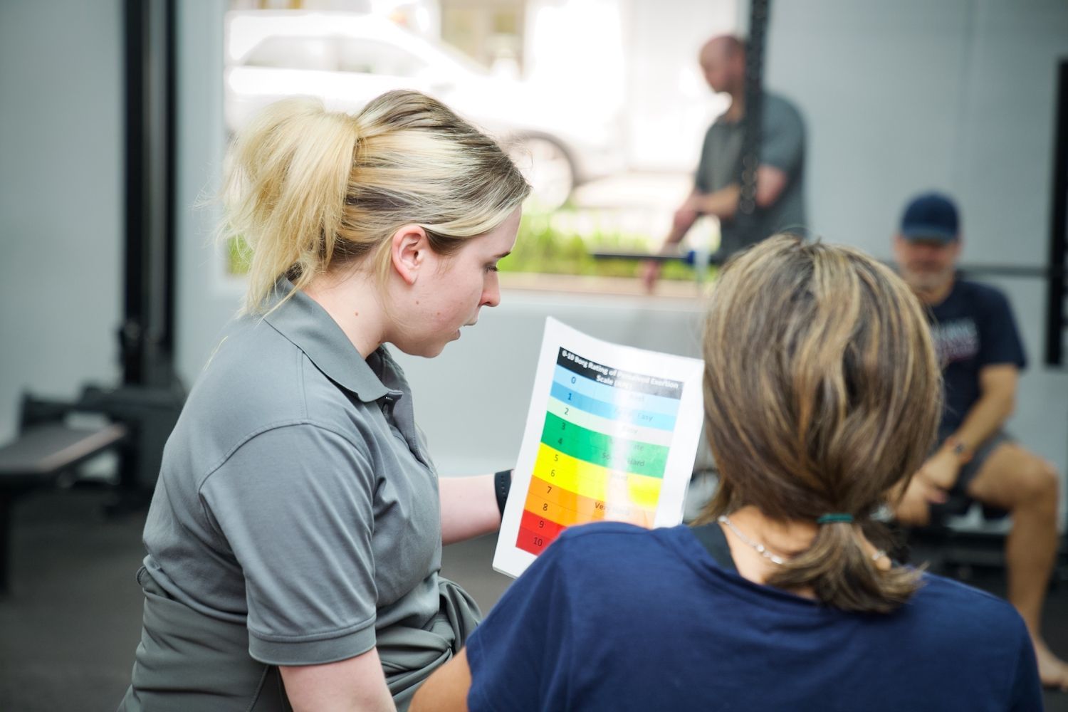
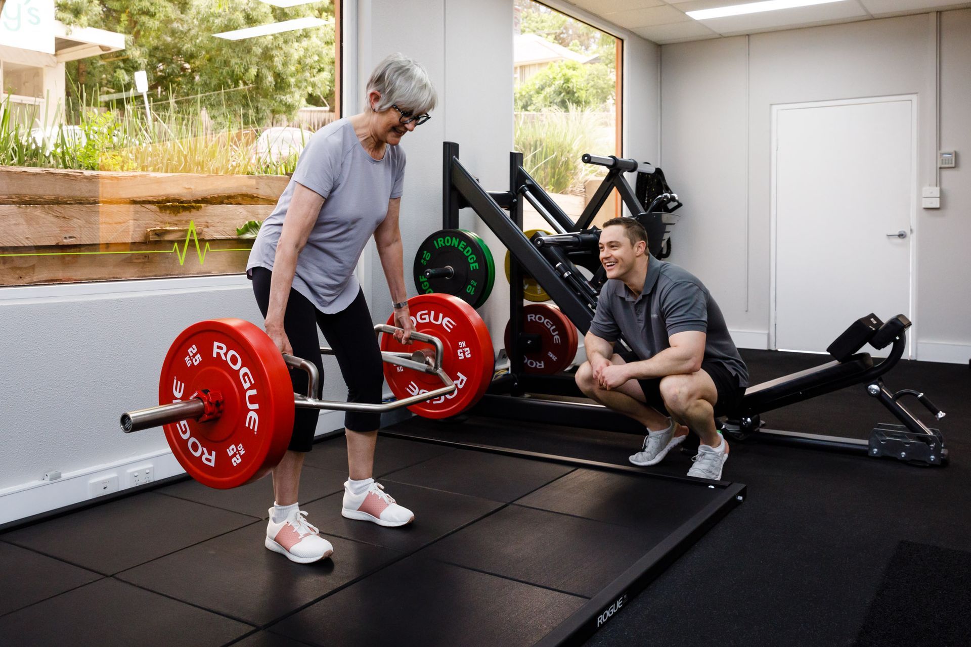
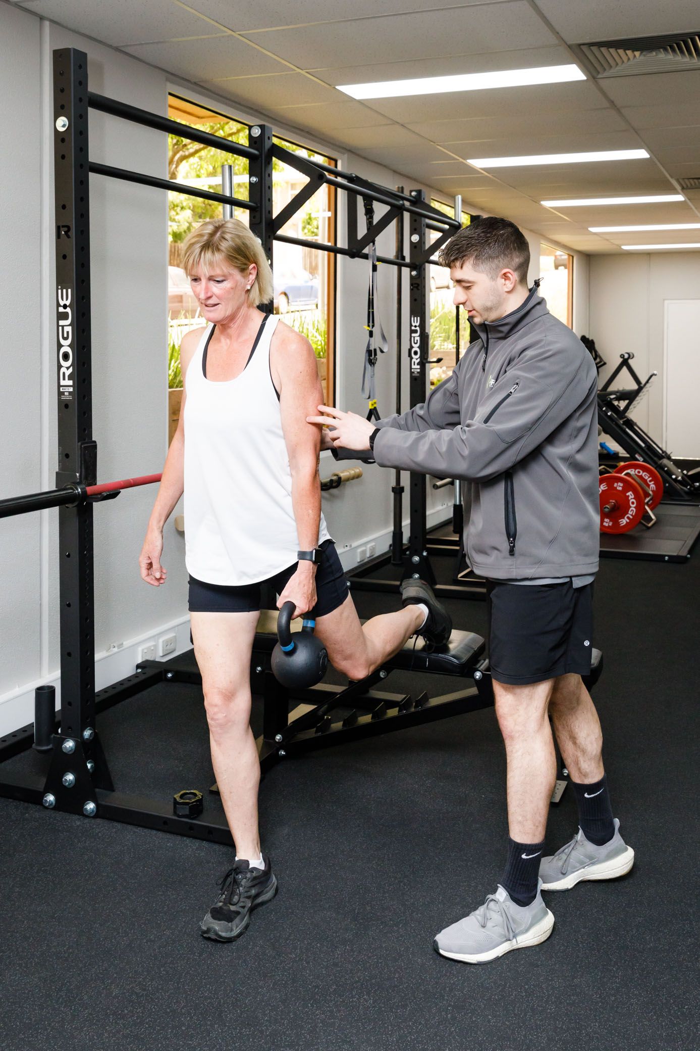
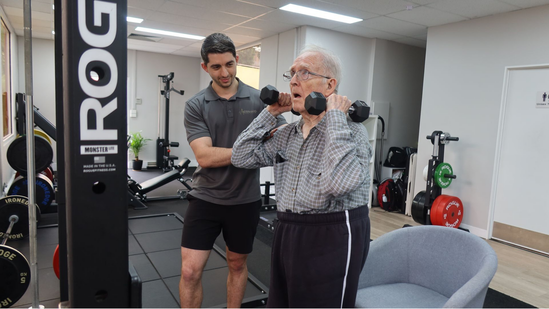
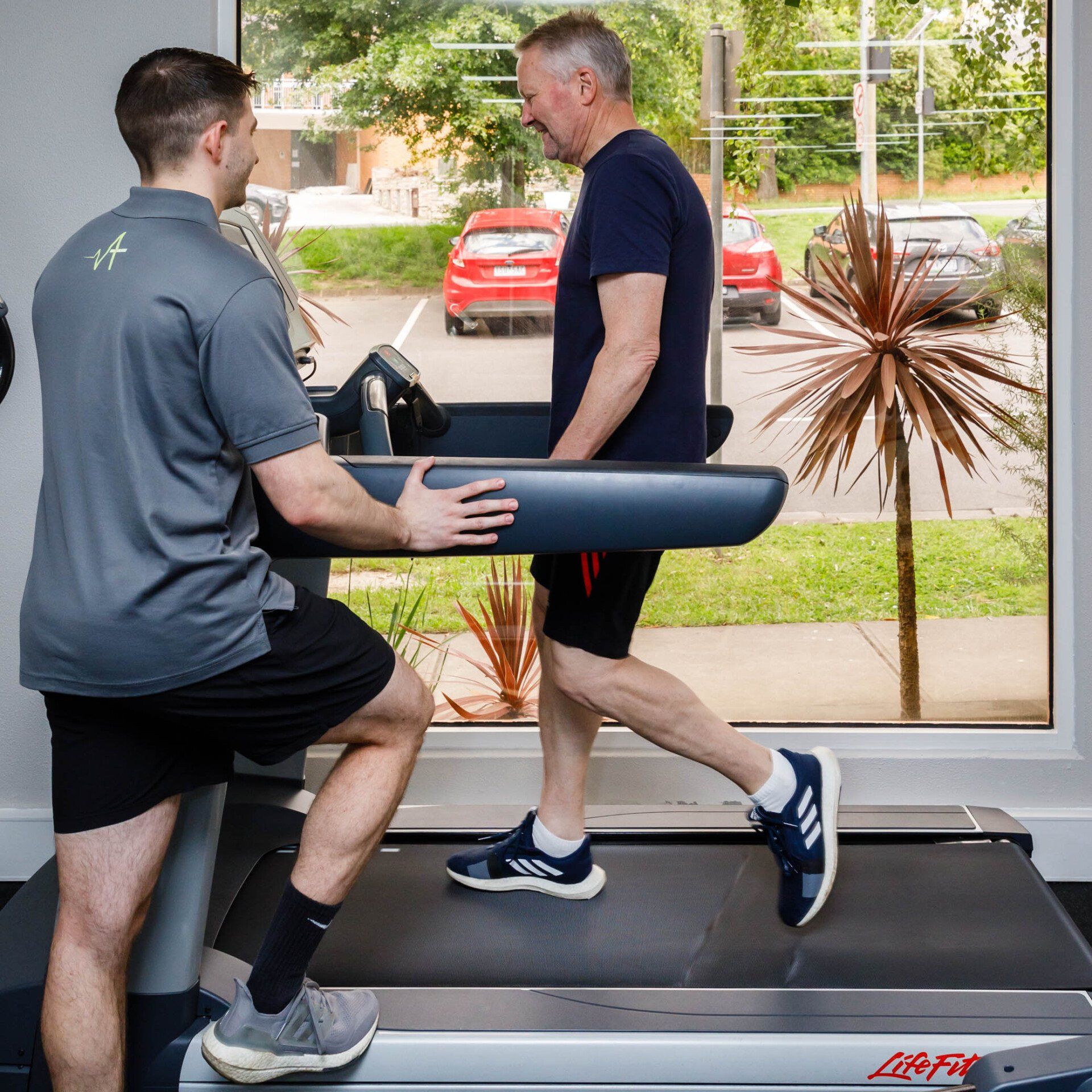
What Exercises Help With Sciatica?
The most effective exercises for sciatica are highly individualised.
Because each person experiences different symptoms, different aggravators, and different levels of nerve sensitivity, what works for one person may not work for another. This is why guided care from a trained professional is invaluable.
A good starting point:
Begin with movements that don’t irritate the nerve but still encourage gentle mobility and load through the spine. Although exercise routines should be individualised for each person, some exercises suitable for the initial stages of sciatica may include:
- Side bends
- Trunk rotations
- Glute bridges
- Leg swings
- Walking
These exercises apply load away from the irritated nerve root, reducing mechanical tension while still promoting healthy movement and mild loading through the spine. Over time, the goal is to gradually build tolerance and restore strength and flexibility without provoking symptoms.
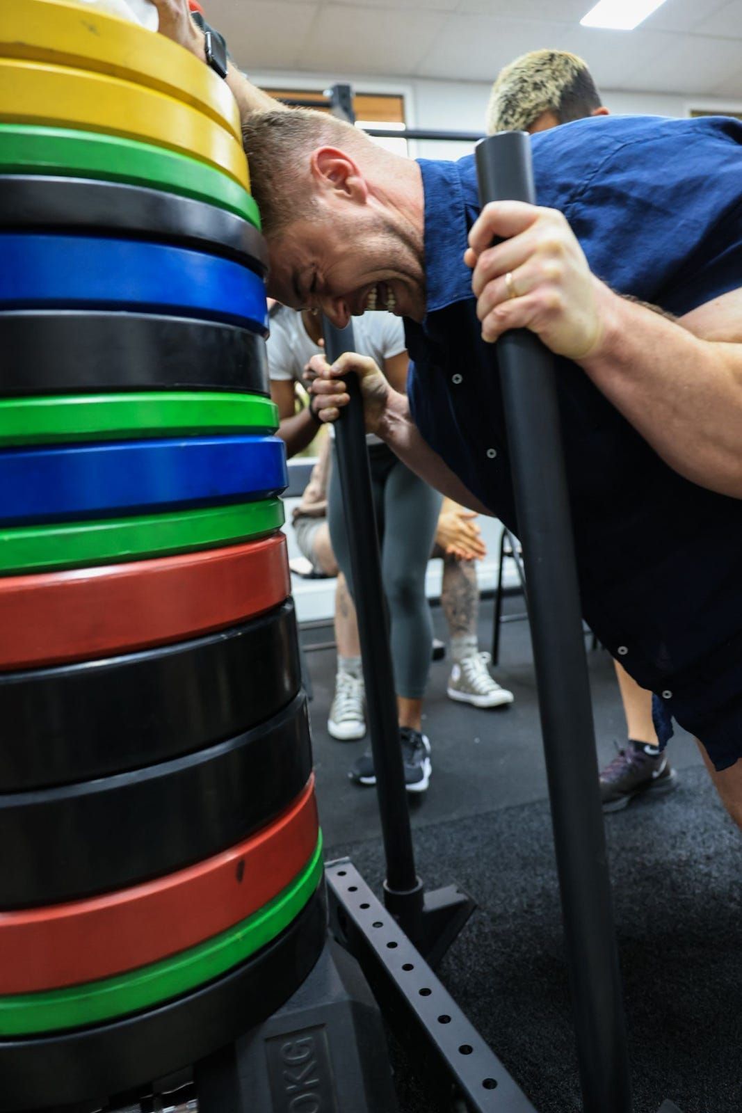
As symptoms settle, exercises must be progressed to include more direct loading, strength work, always tailored to the individual.
Final Thoughts
Sciatica is a specific type of nerve-related leg pain, not just any leg pain that accompanies a sore back. Although it can be intense and limiting, most cases improve significantly with conservative management, movement, and time.
Understanding the signs, causes, and treatment options helps you navigate the condition confidently and choose the right path forward.
References:
Deyo, R. A., & Mirza, S. K. (2016). Herniated Lumbar Intervertebral Disk. The New England Journal of Medicine, 374(18), 1763-1772.
Patel, A. A., Spiker, W. R., Daubs, M., Brodke, D., & Cannon-Albright, L. A. (2011). Evidence for an inherited predisposition to lumbar disc disease. The Journal of Bone & Joint Surgery. American Volume, 93(3), 225–229.
Battié, M. C., Videman, T., Kaprio, J., Gibbons, L. E., Gill, K., Manninen, H., Saarela, J., & Peltonen, L. (2009). The Twin Spine Study: Contributions to a changing view of disc degeneration. The Spine Journal, 9(1), 47–59.
Swain, S., Srikandarajah, S., Haines, T., & Hancock, M. (2020). Strength and exercise-based interventions for knee osteoarthritis: A systematic review and meta-analysis. Journal of Orthopaedic & Sports Physical Therapy, 50(8), 460–478.
Saraceni, N., Kent, P., Ng, L., Campbell, A., & Straker, L. (2021). What kinds of exercise improve pain and physical function in people with knee osteoarthritis? A systematic review and component network meta-analysis. British Journal of Sports Medicine, 56(21), 1228–1239.